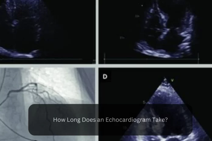
An echocardiogram, a non-invasive medical procedure used to assess the heart, typically takes between 30 minutes to an hour. The duration can vary based on factors like the type of echocardiogram and the complexity of the case. In this article, we delve into the factors influencing the duration of an echocardiogram and provide further insights into the procedure.
What is an Echocardiogram?
An echocardiogram, also known as an echo, is a visual representation of the heart’s movement. During this test, a healthcare provider utilises a handheld wand placed on the chest to capture ultrasound images of the heart’s valves and chambers. These images aid in assessing the heart’s pumping action.
To evaluate blood flow across the heart’s valves, providers often combine echo with Doppler ultrasound and colour Doppler techniques.
One significant advantage of echocardiography is that it does not involve the use of radiation, setting it apart from tests such as X-rays and CT scans that emit small amounts of radiation.
Different- Different types of echocardiogram?
There are various types of echocardiograms, each serving specific purposes in the diagnosis and management of heart disease. These include:
Transthoracic echocardiogram:
This type of echocardiogram involves placing a handheld wand on the chest to obtain images of the heart. It is a common and non-invasive procedure.
Transesophageal echocardiogram:
In this type of echocardiogram, a specialized probe is inserted into the esophagus to capture detailed images of the heart. It provides a closer and clearer view of the heart structures.
Exercise stress echocardiogram:
During this echocardiogram, images of the heart are taken before and after exercise, typically on a treadmill or stationary bike. It helps evaluate the heart’s function and response to physical exertion.
How long does an echocardiogram take?
On average, an echocardiogram typically lasts between 40 to 60 minutes. However, in the case of a transesophageal echo, the procedure may take up to 90 minutes.
When will I need an echocardiogram?
An echocardiogram is ordered by healthcare providers for various reasons. Some common scenarios in which you may need an echocardiogram include:
Investigating Symptoms:
If you are experiencing symptoms, an echocardiogram helps your healthcare provider gather more information to diagnose a potential problem or eliminate possible causes.
Diagnosing Heart Disease:
When your provider suspects heart disease, an echocardiogram is utilised to diagnose the specific issue and gain further insights into its nature.
Monitoring Existing Conditions:
Regular echocardiogram tests may be required for individuals with pre-existing conditions, such as valve disease, to monitor their condition and assess any changes over time.
Preparing for Surgery or Procedure:
Prior to undergoing surgery or a medical procedure, an echocardiogram may be conducted to evaluate your heart’s health and ensure its readiness for the upcoming intervention.
Assessing Surgical or Procedural Outcomes:
After undergoing a surgery or procedure, your provider may order an echocardiogram to assess the effectiveness and outcome of the intervention.
What Does an Echocardiogram Show?
An echocardiogram has the ability to detect various types of heart disease, including:
-
Congenital heart disease
This refers to heart conditions that are present at birth, such as abnormalities in the heart’s structure or function.
-
Cardiomyopathy
This is a condition that affects the heart muscle, leading to problems with its ability to pump blood effectively.
-
Infective endocarditis
An echocardiogram can identify infections occurring in the chambers or valves of the heart.
-
Pericardial disease
This condition affects the pericardium, the two-layered sac surrounding the heart.
-
Valve disease
Echocardiograms are useful in evaluating abnormalities or dysfunction of the heart valves, which regulate blood flow between different chambers of the heart.
Additionally, an echocardiogram can reveal changes in the heart that may indicate:
Aortic aneurysm:
This refers to the abnormal bulging or widening of the aorta, the major blood vessel that carries oxygenated blood from the heart to the rest of the body.
Blood clots:
Echocardiography can detect the presence of blood clots within the heart chambers, which may have implications for stroke risk or circulation issues.
Cardiac tumour:
The imaging capabilities of an echocardiogram allow for the identification of tumours within the heart, whether they are benign or malignant.
FAQ
Q1: What is the purpose of an echocardiogram?
Ans: An echocardiogram is used to assess the structure and function of the heart.
Q2: How long does an echocardiogram typically take?
Ans: An echocardiogram usually lasts between 40 to 60 minutes.
Q3: What conditions can an echocardiogram detect?
Ans: An echocardiogram can detect heart conditions such as congenital heart disease, cardiomyopathy, valve disease, etc.
Q4: What is the purpose of combining Doppler ultrasound with an echocardiogram?
Ans: Doppler ultrasound is used in conjunction with an echocardiogram to assess blood flow across the heart’s valves and identify any abnormalities.


